| New Photos | Animal News | Animal Sounds | Animal Movies | Upload Photo | Copyright | Korean |
|---|
| Funny Animal Photos | Monsters in Animalia | Wiki Articles Fun Facts about Animals | Links | Home | Mobile A.P.A. |
|---|
| Image Info | Original File Name: Sheep Liver Fluke (Fasciola hepatica).jpg Resolution: 448x336 File Size: 39746 Bytes Date: 2004:05:19 17:28:11 Camera: C4040Z (OLYMPUS OPTICAL CO.,LTD) F number: f/2.3 Exposure: 10/200 sec Focal Length: 158/10 Upload Time: 2007:09:25 20:46:12 | |
| Author | Name (E-mail): Unknown | |
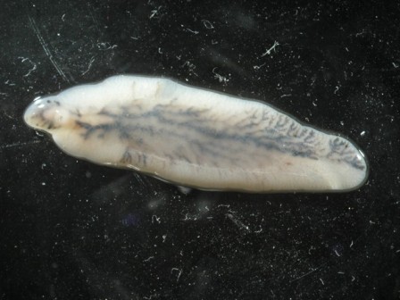
| Subject | Sheep Liver Fluke (Fasciola hepatica) - Wiki | |
 |
| Email : E-Card | Poster | Web Master Delete Edit Info Admin |
| Description | Sheep Liver Fluke (Fasciola hepatica) - Wiki
Fasciola hepatica
Order: Echinostomida Family: Fasciolidae Genus: Fasciola Species: Fasciola hepatica Fasciola hepatica, also known as the common liver fluke or sheep liver fluke, is a parasitic flatworm of the class Trematoda, phylum Platyhelminthes that infects liver of a various mammals, including man. The disease caused by the fluke is called fascioliasis (also known as fasciolosis). F. hepatica is world-wide distributed and causes a great economic losses in sheep and cattle. Life cycle In order to complete its life cycle, F. hepatica requires an aquatic snail as an intermediate host such as Galba truncatula, in which the parasite can reproduce asexually. From the snail, minute cercariae emerge and swim through pools of water in pasture, and encyst as metacercariae on near-by vegetation. From here, the metacercariae are ingested by the ruminant, or in some cases, by humans eating un-cooked foods such as water-cress. Contact with low pH in the stomach causes the early immature juvenile to begin the process of excystment. In the duodenum, the parasite breaks free of the metacercariae and burrows through the intestinal lining into the peritoneal cavity. The newly excysted juvenile does not feed at this stage, but once it finds the liver parenchyma after a period of days, feeding will start. This immature stage in the liver tissue is the pathogenic stage, causing anaemia and clinical signs sometimes observed in infected animals. The parasite browses on liver tissue for a period of up to 5-6 weeks and eventually finds its way to the bile duct where it matures into an adult and begins to produce eggs. Up to 25,000 eggs per day per fluke can be produced, and in a light infection, up to 500,000 eggs per day can be deposited onto pasture by a single sheep. Disease biology In the United Kingdom, Fasciola hepatica is a frequent cause of disease in ruminants - this is most common between March and December. Cattle and sheep are infected when they consume the infectious stage of the parasite from low-lying, marshy pasture. The effects of liver fluke are referred to as fascioliasis, and include anaemia, weight loss and sub-mandibular oedema. Diarrhea is only an occasional consequence of liver fluke. Liver fluke is diagnosed by yellow-white eggs in the faeces. They are not distinguishable from the eggs of Fascioloides magna, although the eggs of F. magna are very rarely passed in sheep, goats or cattle. A serious consequence of the liver damage caused by fascioliasis is that latent Clostridium novyi spores can be activated by the low oxygen conditions in the damaged tracts the parasite forms in the liver - this can lead to "black disease", caused by Clostridium novyi type B or immune-mediated haemolytic anaemia (IMHA) leading to haemoglobinuria caused by Clostridium novyi type D. Treatment The drug of choice in the treatment of fasciolosis is triclabendazole, a member of the benzimidazole family of anthelmintics. The drug works by preventing the polymerisation of the molecule tubulin into the cytoskeletal structures, microtubules. However, resistance of F. hepatica to triclabendazole has already been recorded in Australia and Ireland. http://en.wikipedia.org/wiki/Fasciola_hepatica
| |||
| Copyright Info | AnimmalPicturesArchive.com does not have the copyright for this image. This photograph or artwork is copyright by the photographer or the original artist. If you are to use this photograph, please contact the copyright owner or the poster. |
|
|
|
| |||||||
| CopyLeft © since 1995, Animal Pictures Archive. All rights may be reserved. | ||||||||
Stats