More Protozoa - The Secrets Of Paramecium Caudatum - KLEIN's silver lines Hello again,
here's another issue of my Paramecium caudatum series. As an exception,
it consists of two pictures. That is not because I have not much time
for posting (which is true) but because the second image needs some kind
of explanation which I have to use the first shot for.
If you watch this and remember my previous Paramecium postings the
typical appearance of this guy will already be familiar to you (in case
you missed them all and have no idea what I am talking about please tell
me; I'll repost the previous ones). There is a gorgeous sight of the two
star-shaped contractile vacuoles; the nucleus, being located under the
right vacuole, is also visible. The cut halfway between the contractile
vacuoles is Paramecium's mouth.
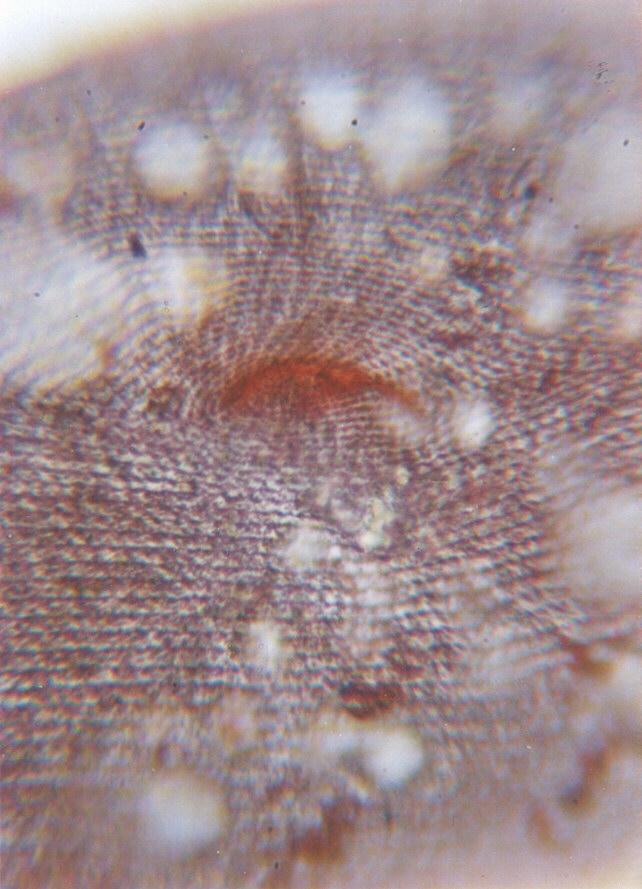
That is exactly what you see in the second shot. It is a close-up of
Paramecium's mouth region; if you watch it on your screen the
magnification is about 4.000 times. That is why the shot doesn't look
particularly crisp; in light microscopy any magnification exceeding
1.200 times goes along with a loss of resolution; it is the light
itself, more precisely its wavelength, which is responsible for that
limitation.
The brownish colour of parts of the shot has a special meaning. I
already told you that I usually prefer watching Protozoa alive, without
the influence of artificial colourings affecting the organism. The
colouring used in this picture, however, reflects one of the most
sensational discoveries in Protozoology.
If you impregnate ciliates with silver salts under certain circumstances
you will be able to see a sort of grid system. This has been renowned
as KLEIN's silver line system according to its discoverer. Most
surprisingly, that grid system not only corresponds with the vital
organs of the ciliate's body but also changes with the situation the
organism has been in before the silver treatment. It has therefore been
interpreted as a primitive nerval system of the ciliates probably
supplying electrical connexions between their organs.
The shot shown here displays the results of a silver nitrate
impregnation as described above applied to Paramecium caudatum. You can
see that the silver lines are located in rows all over the body; in
Paramecium's case almost all of them represent cilia. It becomes clear
that the mouth region is surrounded by concentric rows of cilia; these
produce a vortex which flushes the bacteria right into Paramecium's
mouth.
Thanks for your patience; this was plenty of text and Kilobytes. More
pics to come, but I suppose with a bit less text attached to them.
So long,
Ralf
Content-Type: image/jpeg; name="Paramecium4.jpg"
Content-Type: image/jpeg; name="Silverlines.jpg"
