Protozoa - new scans, #1 - can you stand one more Paramecium caudatum? Hi everybody,
this is my first one in a series of new Protozoa scans. I have about 15
shots left that are worth the post; I shall bring you one a day, maybe
one every two days, dependent on my spare time. At the moment I have no
time to get to the microscope again; I shall hopefully resume
microphotography in autumn or winter, probably in connexion with buying
a digital camera.
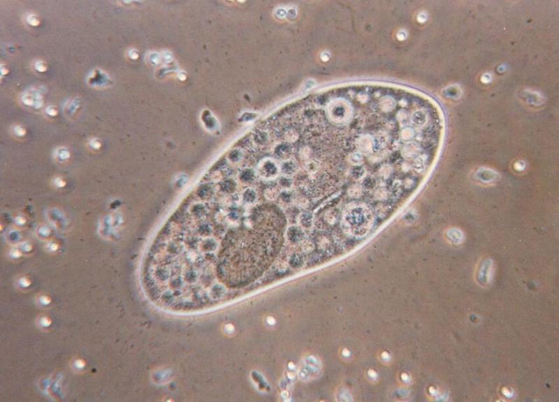
This, now, is my first in a series of about 15 new scans. It features
Paramecium caudatum which may be familiar to you if you have been
watching my first series of Protozoa scans. This Paramecium is swimming
in a sea of food; the small organisms around it are smaller members of
Paramecium's family which is called ciliates or ciliata. The photo shows
nicely that the illumination method being used - phase contrast - does
an extraordinary job in enhancing contrasts: The grey scales of the
image as well as the halos around the object are neither natural nor
chemically added; phase contrast just uses the physical phenomenon of
interference to create visible contrasts from invisible changes in
optical density. The dark-grey spheres inside Paramecium's body are, by
the way, the same organisms which brightly shine like little stars
around Paramecium. It is typical to phase contrast microscopy that the
depth of grey colour of an organism depends on the refraction index of
its surrounding rather than on its own light absorption. The nucleus
also shows up in dark-grey in the lower left of Paramecium; the cut
right to the nucleus is the mouth munching no end of yummy prey items in
this shot.
Enjoy the weekend,
Ralf
name="Paramecium5.jpg"
